Brain: Structure And Function
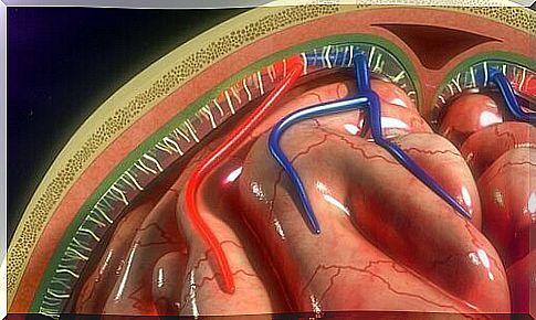
The brain and spinal cord are surrounded by three layers of membranes: meninges. The three meninges are dura mater, arachnoid mater and pia mater. The last two constitute the lepto membranes. Dura mater is also called the pachymeninx.
The most important function of the meninges is to protect the brain. This is a very vulnerable body that needs special protection. No other body needs this, at least not in the same way. In addition, these protective layers are part of the blood-brain barrier.
The meninges originate from another layer known as the primitive meninx, which is composed of elements derived from the mesenchyme and neural crest. The primitive meninx is separated into two different layers: the inner (endomeninx) and the external (ectomeninx).
Endomeninx is different from the arachnoid feeder and the pia feeder and it originates from both mesoderm and ectoderm. On the other hand, the ectomeninx forms the dura mater and the bones of the neurocranium and consists only of the mesoderm.
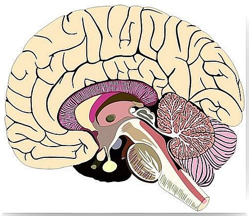
Brains and their structure
Dura mater
The dura mater is the outermost layer of the meninges and it has two layers. The outer layer is the periosteum and contains blood vessels and nerves. It adheres to the inner surface of the skull, in points specially aligned to the base of the skull.
The name of the deepest layer of the dura mater is the meningeal layer. It is responsible for the reflexes that divide the brain into space.
Among these spaces, the most prominent are: Falx cerebri and tentorium cerebelli. In addition, there is no marked boundary between the dura mater and the periosteum. The layers differ histologically in that the meningeal layer has fewer fibroblasts and relatively less collagen (2).
Arachnoid mater
Arachnoid is the middle layer of meninges. It contains the subarachnoid space, which in turn stores cerebrospinal fluid (CSF). The depth of the subarachnoid space varies depending on the ratio of arachnoid mater to pia mater.
Two separate cell layers form the arachnoid artery. The arachnoid barrier cell layer is located after the edge of the cells in the dura mater (3). This layer is filled with cells closely connected by numerous desmosomes and dense compounds. Thus, this layer prevents liquid from moving through it.
The reticular arachnoid layer is deep inside the arachnoid feeder. Its cells connect the subarachnoid space and attach to the pia mater. They also contain the blood vessels that pass through the layer (1).
Arachnoid villi are microscopic structures that play an important role in the absorption of CSF. But the way they work is unclear. Some believe that arachnoid villi may also play a role in regulating CSF volume.
Pia mater
Pia mater is the innermost layer of meninges. It is a fragile vascular structure that surrounds and adheres to the surface of the brain and spinal cord and protects it.
It creates a continuous layer of cells, which are attached to the surface of the brain and follow down into the furrows and depressions in the brain. Desmosomes and interstitial compounds connect the cells, allowing the layer to act as a barrier.
Virchow-Robin rum
Virchow-Robin space is located around the vessels and surrounds the small arteries and arterioles. They pierce the surface of the brain and extend inward from the subarachnoid space (1).
These spaces increase in size as they age, apparently without an associated loss of cognitive function (4). In addition, the expansion of these spaces is related to conditions such as high blood pressure, neuropsychiatric disorders, multiple sclerosis, and trauma (5).
In conclusion, the authors, Patel and Kirmi (2009), emphasized the importance of knowledge about the meninges. It is important to understand their structure, functions and anatomy because it will allow us to understand the disorders related to the meninges.